Home
/ Anatomy Of Musckes Sndctendons / Muscle And Tendon Characteristics Classic Human Anatomy In Motion The Artist S Guide To The Dynamics Of Figure Drawing
Anatomy Of Musckes Sndctendons / Muscle And Tendon Characteristics Classic Human Anatomy In Motion The Artist S Guide To The Dynamics Of Figure Drawing
Anatomy Of Musckes Sndctendons / Muscle And Tendon Characteristics Classic Human Anatomy In Motion The Artist S Guide To The Dynamics Of Figure Drawing. The tightening and relaxing of the calf muscles enables the ankle to bend downward and upward. The muscles, bones, ligaments, and tendons in the back can all be injured and cause back pain. The knee joint is most significantly affected by two major muscle groups: Tendons attach the muscles to each other. The human hand is made up of the wrist, palm, and fingers and consists of 27 bones, 27 joints, 34 muscles, over 100 ligaments and tendons, and many blood vessels and nerves.
As these muscles contract and relax, they move skeletal bones to create movement of the body. Tendons are the reason a muscle can move the bones in our body when muscles contract. It also helps you raise and rotate your arm. The muscles of the upper limb can be divided into 6 different regions: Lying exposed between the protective bones of the superiorly located ribs and the inferiorly located pelvic girdle, the muscles of this region play a critical role in protecting the.
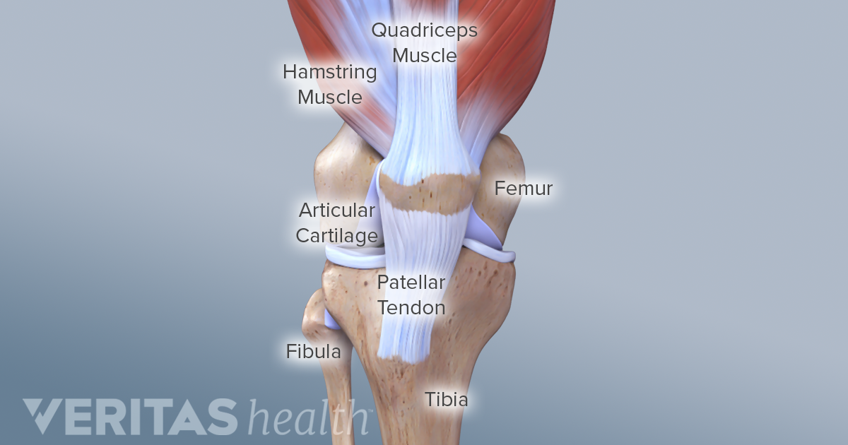
Knee Anatomy from embed.widencdn.net The calf muscles (gastrocnemius and soleus), which are connected to the calcaneus via the achilles tendon. Muscles, either individually or in groups, are supported by fascia. Tendons are the connective tissues that transmit the mechanical force of muscle contraction to the bones; The majority of muscles in the leg are considered long muscles, in that they stretch great distances. Tendons are thick bands of tissue that connect muscles to bones. Lesson on the anatomy of the forearm: Human muscle system, the muscles of the human body that work the skeletal system, that are under voluntary control, and that are concerned with movement, posture, and balance. Extensor carpi radialis brevis extensor carpi radialis longus
Tendons are the reason a muscle can move the bones in our body when muscles contract.
Most structures in the foot are fairly superficial and can be easily palpated. This is lesson 1 on the anatomy of the forearm. As these muscles contract and relax, they move skeletal bones to create movement of the body. By connecting our rigid bones to our powerful muscles, tendons allow us to move. The knee joint is most significantly affected by two major muscle groups: It is comprised of two bones: This is a table of skeletal muscles of the human anatomy. Originates from the bottom of the heel bone and passes underneath the foot to the middle of the sole where it splits into four foot tendons which attach. These muscles are similar to the thenar muscles in both name and organisation. On the other hand, the insertion is where a tendon attaches that muscle to the *more* movable bone. Tendons are thick bands of tissue that connect muscles to bones. The muscles around the knee help to keep the knee stable, well aligned, and moving. There are two main muscle groups around the knee:
Lesson on the anatomy of the forearm: In this lesson, we look at the muscle. Broadly considered, human muscle—like the muscles of all vertebrates—is often divided into striated muscle, smooth muscle, and cardiac muscle. The hip joint is the junction where the hip joins the leg to the trunk of the body. There are two main muscle groups around the knee:
Intramuscular Hamstring Tendon Injury Prognosis Surgical Repair And Rehabilitation from www.sportsinjurybulletin.com The tendon is firmly connected to muscle fibres at one end and to components of the bone at its other end. There are 4 muscles of the pectoral region: Human muscle system, the muscles of the human body that work the skeletal system, that are under voluntary control, and that are concerned with movement, posture, and balance. Pectoralis major, pectoralis minor, serratus anterior and subclavius.collectively, these muscles are involved in movement and stabilisation of the scapula, as well as movements of the upper limb. Extensor carpi radialis brevis extensor carpi radialis longus The smaller bone that runs alongside the tibia (fibula) and the kneecap (patella) are the other bones that make the knee joint. The thigh bone or femur and the pelvis which is made up of three bones called ilium, ischium, and pubis. Tendons attach the muscles to each other.
The quadriceps are a collection of 4 muscles on the front of the thigh and are responsible for straightening the knee by bringing a bent knee to a straightened position.
The muscles around the knee help to keep the knee stable, well aligned, and moving. The thigh bone or femur and the pelvis which is made up of three bones called ilium, ischium, and pubis. There are two sets of flexor foot tendons, each made up of two muscles and tendons, one set that bends the big toe, the other set that bends the remaining four toes. Most structures in the foot are fairly superficial and can be easily palpated. Muscles and tendons of the leg, find out more about muscles and tendons of the leg. Pectoral, shoulder, upper arm, anterior forearm, posterior forearm, and the hand. Movement occurs when our muscles pull on our bones, relocating them. Muscles, either individually or in groups, are supported by fascia. When the muscle contracts, the tendons are pulled, and the bone is moved. A solid understanding of anatomy is essential to effectively diagnose and treat patients with foot and ankle problems. Originates from the bottom of the heel bone and passes underneath the foot to the middle of the sole where it splits into four foot tendons which attach. In this lesson, we look at the muscle. It also helps you raise and rotate your arm.
There are 4 muscles of the pectoral region: The muscles you probably know the best are your. This is lesson 1 on the anatomy of the forearm. Pectoral, shoulder, upper arm, anterior forearm, posterior forearm, and the hand. Tendons are thick bands of tissue that connect muscles to bones.
Calf Muscle Tightness Achilles Tendon Length And Lower Leg Injury Mountain Peak Fitness from static1.squarespace.com Most of the muscles which act on the wrist joint are situated within the forearm, with only the tendon crossing the joint and inserting on the hand. Tendon, tissue that attaches a muscle to other body parts, usually bones. The calf muscles (gastrocnemius and soleus), which are connected to the calcaneus via the achilles tendon. Human muscle system, the muscles of the human body that work the skeletal system, that are under voluntary control, and that are concerned with movement, posture, and balance. The quadriceps muscles provide strength and power with knee extension (straightening). Every skeletal muscle has three main parts: Muscles, either individually or in groups, are supported by fascia. In this lesson, we look at the muscle.
Every skeletal muscle has three main parts:
The muscles, bones, ligaments, and tendons in the back can all be injured and cause back pain. These muscles are similar to the thenar muscles in both name and organisation. Similar to ligaments, they are made of collagen and can withstand increased tension. The tendon is firmly connected to muscle fibres at one end and to components of the bone at its other end. A tendon connects the muscle to the bone. Human anatomy for muscle, reproductive, and skeleton. Every skeletal muscle has three main parts: On the other hand, the insertion is where a tendon attaches that muscle to the *more* movable bone. Anatomy is a road map. Pectoralis major, pectoralis minor, serratus anterior and subclavius.collectively, these muscles are involved in movement and stabilisation of the scapula, as well as movements of the upper limb. The muscles of the abdomen, lower back, and pelvis are separated from those of the chest by the muscular wall of the diaphragm, the critical breathing muscle. This is lesson 1 on the anatomy of the forearm. The quadriceps are a collection of 4 muscles on the front of the thigh and are responsible for straightening the knee by bringing a bent knee to a straightened position.
Share :
Post a Comment
for "Anatomy Of Musckes Sndctendons / Muscle And Tendon Characteristics Classic Human Anatomy In Motion The Artist S Guide To The Dynamics Of Figure Drawing"


Post a Comment for "Anatomy Of Musckes Sndctendons / Muscle And Tendon Characteristics Classic Human Anatomy In Motion The Artist S Guide To The Dynamics Of Figure Drawing"